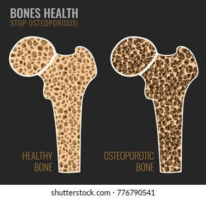
Bone Cross Section : Bone Cross Section Images Stock Photos Vectors Shutterstock. (2) cross‐sectional moment of inertia (csmi): Browse 53 bone marrow cross section stock photos and images available, or search for bone cross section or bone cells to find more great stock photos and pictures. As the names suggest compact bone looks compact and the spongy bone looks like sponges. Thin sections are used for microradiography and for observation with transmitted light. While it is not as hard as compact bone, spongy bone plays an important role of protecting the marrow where blood cells are produced.
Vector illustration scheme of bone cross section. As the names suggest compact bone looks compact and the spongy bone looks like sponges. This slide contained a cross section of a very small bone, and you are looking at the entire thickness of the shaft of the bone. Human bone, cross section diagram of femur showing osteon, veins, marrow. Compact bone is the outer layer and the spongy bone forms the inner layer.
Color Illustration Of Cross Section Of Right Arm Superior View Showing Muscles Bone Nerves Arteries And Veins Orthopaedic Surgical Anatomy Teaching Collection Usc Libraries Digital Collections from digitallibrary.usc.edu Compact bone is the outer layer and the spongy bone forms the inner layer. Explaned distal and proximal epiphysis. At the end of the bone is the epiphysis, which in young people is separated from the. A property of the cross‐sectional area that represents the magnitude of the greatest bending rigidity of the section (cm 4). Muscles and bones of the human body 12 photos of the muscles and bones of the human body anatomy bones of the human body quiz, major muscles and bones in the human body, muscles and bones in the human body, number of muscles and bones in the human body. Internal structure of a human long bone, with a magnified cross section of the interior. Table 1 describes the bone markings, which are illustrated in (figure 4). Learn vocabulary, terms, and more with flashcards, games, and other study tools.
Table 1 describes the bone markings, which are illustrated in (figure 4).
The upper (biting) surfaces of the tooth are at top, with the lower sections (bottom) embedded in the gums and jaw bone (not shown). Compact bone is the outer layer and the spongy bone forms the inner layer. By printing out this quiz and taking it with pen and paper creates for a good variation to only playing it online. The compact bone is made up of osteon. The surface features of bones vary considerably, depending on the function and location in the body. This is an online quiz called bone cross section. Muscles and bones of the human body 12 photos of the muscles and bones of the human body anatomy bones of the human body quiz, major muscles and bones in the human body, muscles and bones in the human body, number of muscles and bones in the human body. Red bone marrow fills the spaces between the spongy bone in some long bones. There is a printable worksheet available for download here so you can take the quiz with pen and paper. A cross section of a human long bone. Compact bone is very different from the other tissues you have seen. Cross‐sectional area is derived from the integral of the bone mass profile across the narrow region. Related posts of cross section of human bone diagram muscles and bones of the human body.
Two types of bone tissues in cross section of a long bone : Bone structure right foot 12 photos of the bone structure right foot bone structure in. Compact bone is the outer layer and the spongy bone forms the inner layer. Table 1 describes the bone markings, which are illustrated in (figure 4). Start studying bone cross sections.
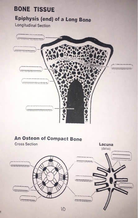
Solved Bone Tissue Epiphysis End Of A Long Bone Longitu Chegg Com from media.cheggcdn.com There is a printable worksheet available for download here so you can take the quiz with pen and paper. Related posts of cross section of a long bone bone structure right foot. I am not an expert on this subject, so i was wondering if anyone could put their input on this image. The two types of bone marrow are red bone marrow, known as myeloid tissue, and yellow bone marrow, or fatty tissue. Start studying bone cross section 2. Thin sections are used for microradiography and for observation with transmitted light. The central tubular region of the bone, called the diaphysis, flares outward near the end to form the metaphysis, which contains a largely cancellous, or spongy, interior. Compact bone is the outer layer and the spongy bone forms the inner layer.
Table 1 describes the bone markings, which are illustrated in (figure 4).
It consists of two layers; While it is not as hard as compact bone, spongy bone plays an important role of protecting the marrow where blood cells are produced. The upper (biting) surfaces of the tooth are at top, with the lower sections (bottom) embedded in the gums and jaw bone (not shown). The central tubular region of the bone, called the diaphysis, flares outward near the end to form the metaphysis, which contains a largely cancellous, or spongy, interior. The compact bone is made up of osteon. There is a printable worksheet available for download here so you can take the quiz with pen and paper. Find the perfect cross section of bone stock photos and editorial news pictures from getty images. The surface features of bones vary considerably, depending on the function and location in the body. This is known as the periosteum. An outer 'fibrous layer' containing mainly fibroblasts, and an inner 'cambium layer' containing progenitor cells. Product is not alive nor is it edible. At the end of the bone is the epiphysis, which in young people is separated from the. Red bone marrow fills the spaces between the spongy bone in some long bones.
Examination of bone and tissue samples bones and tissues are studied by two different methods. Table 1 describes the bone markings, which are illustrated in (figure 4). Internal structure of a human long bone, with a magnified cross section of the interior. Learn vocabulary, terms, and more with flashcards, games, and other study tools. 100x first focus in the compact decalcified bone (cb) on the left part of the image, you can see small dots, which are.
Bone Cross Section Images Stock Photos Vectors Shutterstock from image.shutterstock.com To the left is muscle tissue, and to the right is bone marrow. The surface features of bones vary considerably, depending on the function and location in the body. The compact bone is made up of osteon. Select from premium cross section of bone of the highest quality. An outer 'fibrous layer' containing mainly fibroblasts, and an inner 'cambium layer' containing progenitor cells. Explaned distal and proximal epiphysis. Human bone, cross section diagram of femur showing osteon, veins, marrow. Compact bone is very different from the other tissues you have seen.
Browse 9,121 bone cross section stock photos and images available, or search for bone marrow or bone structure to find more great stock photos and pictures.
Muscles and bones of the human body 12 photos of the muscles and bones of the human body anatomy bones of the human body quiz, major muscles and bones in the human body, muscles and bones in the human body, number of muscles and bones in the human body. Browse 53 bone marrow cross section stock photos and images available, or search for bone cross section or bone cells to find more great stock photos and pictures. Human bone, cross section diagram of femur showing osteon, veins, marrow. Internal structure of a human long bone, with a magnified cross section of the interior. There is a printable worksheet available for download here so you can take the quiz with pen and paper. There are three general classes of bone. This is a short tutorial using blender 2.8 that shows how to create a bone cross section and using images to create the textures.hope you enjoy and please su. Bone structure right foot 12 photos of the bone structure right foot bone structure in. The surface features of bones vary considerably, depending on the function and location in the body. Two types of bone tissues in cross section of a long bone : Start studying bone cross section 2. The wider section at each end of the bone is called the epiphysis (plural = epiphyses), which is filled internally with spongy bone, another type of osseous tissue. (2) cross‐sectional moment of inertia (csmi):
Share :
Post a Comment
for "Bone Cross Section : Bone Cross Section Images Stock Photos Vectors Shutterstock"


Post a Comment for "Bone Cross Section : Bone Cross Section Images Stock Photos Vectors Shutterstock"